Western Sydney University researchers have successfully developed a revolutionary way to increase the lifespan of live tissue required for scientific and medical research by 400 per cent.
Previously, the viability of live tissue was only six hours, which meant research time was limited. Using this new technology, live tissue is still viable after 24 hours.
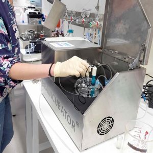
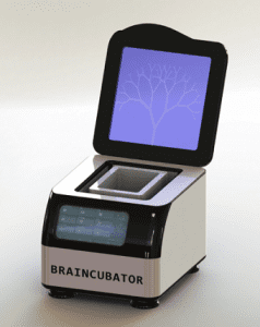
Dr Yossi Buskila and Dr Paul Breen from the MARCS Institute for Brain, Behaviour and Development built the Braincubator to increase the testing and research capabilities of live tissue, and also drastically reduce the number of animals sacrificed for research purposes, namely rats and mice.
Because rats share similar DNA structures with humans, and often suffer from the same diseases, they are commonly used in medical research.
Dr Buskila, who is a neuroscientist, uses brain slices from rats to study the way neurons communicate and process electrophysiological signals.
“In vitro brain slice preparations are instrumental in developing our understanding of the nervous system, but previously the lifespan of an acute brain slice was limited to around six hours,’’ he said.
“This reduces potential experimentation time and leads to considerable waste of neural tissue.
“My aim was to provide a practical and feasible solution that harnessed opportunities where rare live tissue was obtained for testing and research purposes – and to use these tissues in the most effective and ethical way.”
Dr Buskila said the main reason for the short lifespan of live tissue was its susceptibility to bacterial and environmental degradation which caused the cells to die.
He said the Braincubator was so effective because it acted as an incubation system – thus its name – that prevented the growth of bacteria, slowing down cell deterioration.
“Brain slices require an environment that simulates their natural environment in order to maintain cellular metabolic activity and electrophysiological function,” he said.
“The Braincubator – which is now a registered trade mark – works by controlling the temperature and pH levels of the artificial cerebral spinal fluid in which the tissue is incubated.
“This system is effective at maintaining extremely low bacterial levels by continually passing this artificial fluid through a UVC filtration system.”
Dr Buskila said a few attempts had been made over the years to maximize cellular longevity, however, none as successful as this concept.
“This technology has already been used to extend viability of retinal tissue for research, and we are also exploring options to expand it to other tissues such as spinal cord and organ tissue,” he said.
“I believe the use of the Braincubator will have far-reaching benefits for the efficiency of research by effectively quadrupling the amount of data that can be produced from one animal through extending live tissue sustainability.”